The non-phospho-XCR1 receptor antibody is directed against the distal part of the carboxyl-terminal tail of human XCR1. It can be used to detect total XCR1 receptors in Western blots independent of phosphorylation. The non-phospho-XCR1 antibody can also be used to isolate and enrich XCR1 receptors from cell and tissue lysates. It also detects XCR1 in cultured cells and tissue sections by immunohistochemistry.
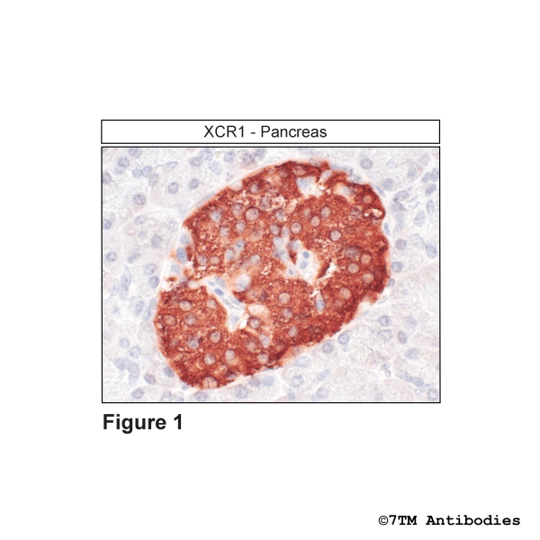
Figure 1. Immunohistochemical identification of XCR Chemokine Receptor 1 in pancreas. Sections were dewaxed, microwaved in citric acid, and incubated with anti-XCR1 (non-phospho-XCR Chemokine Receptor 1) antibody (7TM0075N-IC) at a dilution of 1:100. Sections were then sequentially treated with biotinylated anti-rabbit IgG and avidin-biotin solution. Color was developed by incubation in 3-amino-9-ethylcarbazole (AEC), and sections were counterstained with hematoxylin.
Figure 2. Validation of the XCR Chemokine Receptor 1 in transfected HEK293 cells. Native HEK293 cells (MOCK) or HEK293 cells stably expressing the XCR Chemokine Receptor 1 (XCR1) were lysed and immunoblotted with the phosphorylation-independent anti-XCR1 antibody (7TM0075N-IC) at a dilution of 1:1000.


, XCR Chemokine Receptor 1 Antibody")


, CX3C Chemokine Receptor 1 Antibody")
, CXCR3 Chemokine Receptor Antibody")


